A punch biopsy is a quick and relatively painless procedure to collect a sample of skin tissue from your dog. This sample is then examined by a veterinarian to diagnose various skin conditions.
The procedure typically takes only a few minutes to perform. Your veterinarian will use a special tool to remove a small, circular piece of skin from your dog's body.
The punch biopsy site usually heals quickly, and your dog may experience some mild discomfort or swelling afterwards. However, this is usually temporary and can be managed with medication if necessary.
For your interest: Biopsy on Dog Lump
What Is a Skin Biopsy?
A skin biopsy is a procedure where a small sample of skin is removed and examined under a microscope to determine the nature of a suspicious skin growth or area of skin that has failed to respond to treatment.
Most skin biopsies are performed on a small area of skin, requiring only a few sutures.
A fresh viewpoint: Service Dog Vest for Small Dogs
The area to be biopsied is often numbed with a local anesthetic to minimize discomfort.
In some cases, a sedative or general anesthetic may be necessary, depending on the area involved or the patient's condition.
Either the entire mass or a small section of skin is removed for examination by a veterinary pathologist.
The pathologist will examine the tissues under a microscope to determine the type of cells, their relationship to each other, and any evidence of malignancy.
Preparing for a Biopsy
Timing is everything when it comes to a biopsy. Active disease is required to obtain an accurate diagnosis, so it's best to wait for pustule recurrence if pemphigus foliaceus is suspected.
Corticosteroids and bacterial infections should be withdrawn for several weeks prior to the biopsy to avoid masking the primary pathology. This will give your veterinarian a clear picture of the disease.
Local anesthesia is usually sufficient for a biopsy, but sedation may be necessary to minimize patient movement and increase comfort. A combination of dexmedetomidine and butorphanol can be used for sensitive areas like the nasal planum and paws.
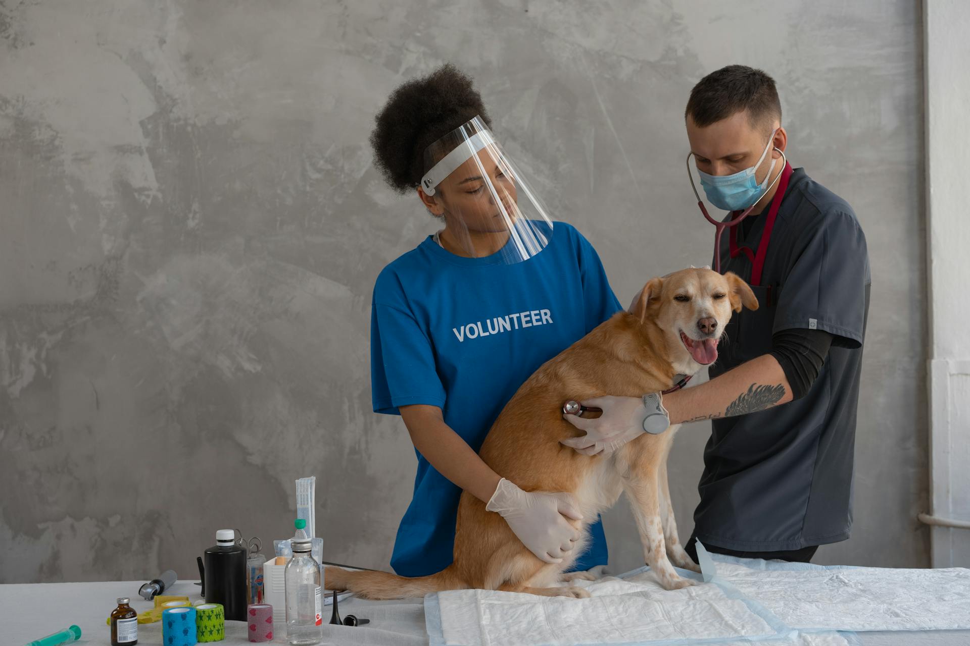
When to Have a Dog's Skin Biopsy
Any suspicious skin lesion on your dog should be biopsied. This includes lumps or masses that appear suddenly or grow rapidly, as these are characteristics associated with malignant skin tumors.
Punch biopsies are not always the best option, especially for vesicles and bullae, as the shearing force can damage them. A punch may also not reach deep enough to obtain a diagnostic sample for diseases of the panniculus.
Any area of poorly healing or abnormal skin should be biopsied. This includes skin that is chronically inflamed or irritated, or has an unusual color or texture.
Incisional elliptical biopsies are preferred for ulcers, vesicles, and bullae, as they allow for the inclusion of unaffected skin for diagnosis. Samples are cut in half longitudinally during processing.
Preparation
Timing is everything when it comes to scheduling a biopsy. Active disease is required to obtain an accurate diagnosis, and some lesions may be transient.
It's essential to wait for pustule recurrence if pemphigus foliaceus is suspected, as this can help ensure an accurate diagnosis. Withdrawal of corticosteroids and treatment of bacterial infections for several weeks prior to taking biopsy samples is usually necessary to avoid masking the primary pathology.
Biopsies can often be performed without sedation, but sedation may be necessary to minimize patient movement and increase patient comfort.
Biopsy Submission
When handling a biopsy sample, it's essential to minimize handling to prevent artifacts that could prevent diagnosis.
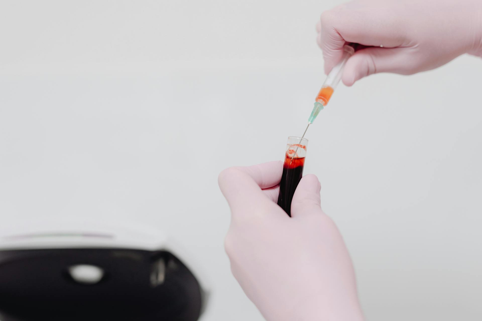
The sample should be placed in 10% neutral buffered formalin within 30 minutes of biopsy.
A ratio of roughly 10 parts formalin to 1 part tissue sample by volume is ideal for preservation.
Small samples, like those from needle-core devices and small biopsy punches, should be placed into plastic cassettes to prevent sample loss.
Submission forms should include the animal's signalment, lesion description, rate of lesion progression, and relevant lab results.
Photos of the lesion before biopsy can be added to the submission form to provide clinical context.
If the pathological diagnosis doesn't fit the clinical history, consider reaching out to the pathologist for additional information or a second opinion.
Biopsy Types and Techniques
A punch biopsy is a common and effective way to collect a sample for diagnosis. This technique is often used for skin lesions.
A 6-mm punch is typically used for a punch biopsy. This size punch is suitable for most skin lesions, but smaller punches are reserved for sensitive areas like the pinnae, nasal planum, or footpads of small dogs and cats.
It's essential to center the punch in the lesion, unless it's an ulcer, and to avoid including too much normal skin. This ensures that the tissue section cut in half at the lab captures the lesion.
Dull punches can cause tissue compression and artifact, so it's best to use a fresh punch for each animal. If the punch dulls during sample collection, switch to a new one to prevent these issues.
Rotating the punch in one direction helps prevent shear artifact, and decreased resistance is felt once the punch reaches the subcutis.
You might enjoy: One Punch Man Dog Hero
Understanding Biopsy Results
A skin biopsy can tell us whether we should be concerned about a lesion, and it will differentiate between infections, allergic skin disease, autoimmune skin disease, and tumors - benign or malignant.
Your veterinarian will use the biopsy results to determine the most appropriate treatment for your dog's condition.
A biopsy provides information about the nature of the abnormality, allowing your veterinarian to determine the prognosis for the condition so that you will know what to expect during the course of the disease.
Skin biopsies are a relatively simple, painless procedure that can speed recovery and improve quality of life for many patients.
To understand biopsy results, it's essential to consider the clinical context, including the animal's signalment, lesion description, rate of lesion progression, and any relevant lab results or imaging findings.
Submission forms should include photos of the lesion before biopsy to provide clinical context and increase the odds of an accurate diagnosis.
Histopathologic results of a tumor biopsy should always be considered within the clinical context, and if the pathological diagnosis doesn't fit the clinical history, it's best to reach out to the pathologist for additional information or a second opinion.
A ratio of roughly 10 parts formalin to 1 part tissue sample by volume is ideal to ensure appropriate preservation of the biopsy sample.
Frequently Asked Questions
How long does it take for a dog to recover from a punch biopsy?
Recovery time for a punch biopsy in dogs typically takes 1-2 days, as the procedure is usually less invasive and anesthesia effects wear off quickly. However, follow-up care and medication instructions should be closely followed as prescribed by your veterinarian.
How much does a punch biopsy cost for a dog?
A punch biopsy for a dog typically costs between $400-$800, depending on the location and veterinarian. This cost may vary, so it's best to consult with a veterinarian for a more accurate estimate.
Featured Images: pexels.com