Medial patellar luxation is a common condition in dogs where the kneecap slips out of place. It's estimated that up to 1 in 5 dogs will experience some degree of medial patellar luxation in their lifetime.
This condition can be caused by a variety of factors, including genetics, obesity, and trauma. Dogs with shallow knee joints or weak quadriceps muscles are more prone to medial patellar luxation.
In some cases, the kneecap may only slip out of place occasionally, while in other cases it may be a chronic problem. The severity of the condition can range from mild to severe, with more severe cases requiring surgery.
Symptoms of medial patellar luxation can include pain, stiffness, and difficulty walking.
What Is Patellar Luxation?
Patellar luxation is a condition where the kneecap, or patella, pops out of place in the stifle joint (knee). This can happen in a medial or lateral position.
The patella is usually reducible, meaning it can be gently pushed back into place. However, laxity of the medial collateral ligament may be evident, and the medial retinacular tissues of the stifle joint are often thickened.
Patellar luxations can be classified into several categories, including medial luxation, lateral luxation, and luxation resulting from trauma. These categories are based on the breed and location of the luxation.
Here are the categories of patellar luxation:
- Medial luxation (toy, miniature, and large breeds)
- Lateral luxation (toy and miniature breeds)
- Lateral luxation (large and giant breeds)
- Luxation resulting from trauma (various breeds)
Animals can be affected by patellar luxation as early as eight weeks of age. The most notable finding is a knock-knee (genu valgum) stance.
Causes and Risk Factors
Medial patellar luxation is a condition that's often considered developmental, meaning it's recognized as the animal grows. This typically occurs when the animal is less than 1 year old.
Trauma can also cause medial patellar luxation, particularly if a bone in the hind limb is broken and not properly realigned.
Abnormalities such as abnormal placement of the quadriceps muscles, abnormal angulation of the head of the femur, and others have been implicated in the cause of medial patellar luxation.
What Causes
Medial patella luxation is often considered a developmental condition, meaning it's recognized as the animal grows, and is usually diagnosed when they're less than 1 year old.
Trauma can also cause patella luxation, especially if a bone is broken in the hind limb and not properly realigned.
Abnormal placement of the quadriceps muscles is one of the abnormalities that can lead to medial patella luxation.
Abnormal angulation of the head of the femur can also contribute to this condition.
Surgery on the stifle joint for other reasons can sometimes result in patella luxation, although this is less common.
In some cases, a previous injury or surgery can increase the risk of developing patella luxation.
Toy, Miniature, and Large Breeds
Toy breeds, weighing less than 7 pounds, have a higher risk of dental problems due to their small jaw size. This can lead to overcrowding and make it difficult to clean their teeth.
Miniature breeds, weighing between 7 and 15 pounds, are more prone to obesity, which increases their risk of various health issues, including diabetes and arthritis.
Large breeds, weighing over 50 pounds, are more susceptible to joint problems, such as hip dysplasia, due to their rapid growth rate and increased body weight.
Clinical Signs and Diagnosis
Medial patellar luxation can be a sneaky condition, especially in small breed dogs like Yorkshire terriers, Chihuahuas, and Toy Poodles. These breeds are often diagnosed with MPL without showing any clinical problems.
In some cases, dogs will limp due to patella luxation, and when lameness is noted, surgical correction is usually indicated. Medical management with anti-inflammatories may be tried initially, but it's generally ineffective and can lead to further complications.
Dogs with medial patella luxation may exhibit a classic "skipping lameness" where the patella pops out, causing the dog to lift the affected pelvic limb and skip on three legs. This is a common symptom in dogs with Grade II patella luxation.
The degree of luxation determines the severity of clinical signs. In Grades 1 and 2, lameness is evident only when the patella is in the luxated position, and the leg is carried with the stifle joint flexed. In contrast, Grade 3 and 4 animals exhibit a crouching, bowlegged stance with the feet turned inward.
Here's a breakdown of the clinical signs associated with medial patellar luxation:
It's essential to note that permanent luxation can render the quadriceps ineffective in extending the stifle, leading to a reduction in the dog's mobility.
Treatment and Surgery
Treatment and surgery are often necessary to correct medial patellar luxation.
The most commonly performed procedures for MPL surgery are tibial tuberosity transposition, deepening of the femoral groove, and soft tissue modification of the joint capsule. These procedures aim to realign the patella and quadriceps, and reestablish straight line force down the pelvic limb.
Surgery can be performed in combination to achieve the best results. The goal is to stabilize the patella in its correct position, making it more difficult for it to luxate.
A combination of four surgical modifications is most commonly used to achieve this goal: trochleoplasty, lateral tibial tuberosity transposition, medial desmotomy, and lateral imbrication.
How Is Treated?
Medial patella luxation treatment is a combination of procedures aimed at re-aligning the patella and quadriceps, and reestablishing straight line force down the pelvic limb. The goal is to make it more difficult for the patella to luxate.
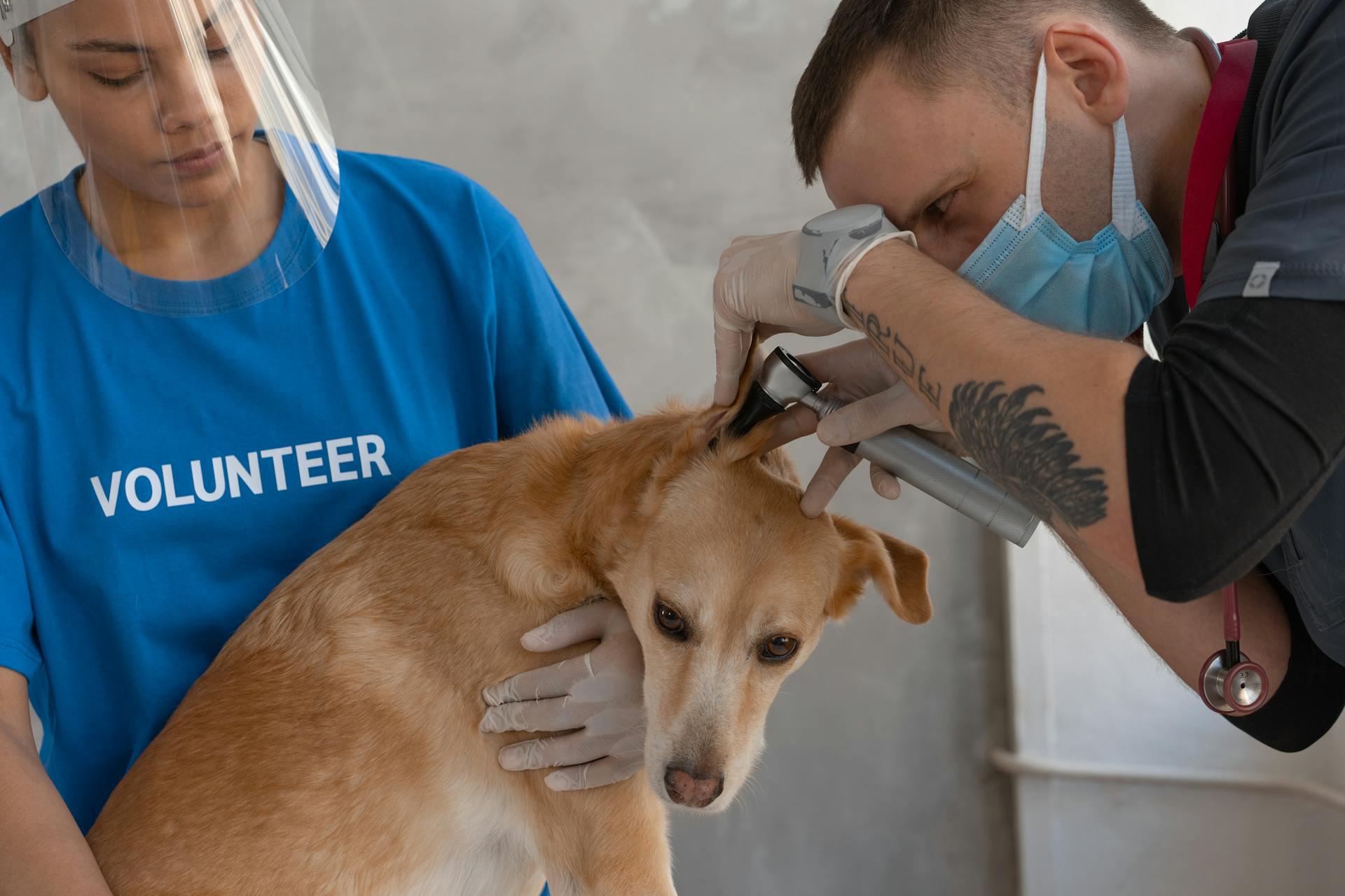
Surgical correction of MPL generally requires several procedures used in combination, including tibial tuberosity transposition, deepening of the femoral groove, and soft tissue modification of the joint capsule.
Tibial tuberosity transposition involves shifting the insertion point of the patellar tendon laterally, re-aligning the patella with its groove. This procedure is often performed in combination with other procedures.
Deepening of the femoral groove, also known as sulcoplasty, is a technique used to deepen a shallow trochlear groove. Three methods are commonly used: sulcoplasty, trochlear wedge recession, and trochlear block recession.
Here are the three methods used to deepen a shallow trochlear groove:
The procedures used in MPL surgery aim to stabilize the patella in its correct position, making it more difficult for the patella to luxate.
Who Can Benefit from MPL Surgery?
MPL surgery can be a game-changer for dogs with certain types of patellar luxations.
Patients with symptomatic grade one luxations and mild grade two luxations can usually be managed successfully with medications, joint health supplements, nutraceuticals, and weight management.
Dogs with grade two luxations that are more severe, as well as all grade three and four luxations, require surgery to restore comfort and limb function.
Here's a breakdown of the different grades of patellar luxations and their corresponding treatment options:
Post-Operative Care Requirements
Physical activity must be restricted until the bones have fully healed. This is crucial to ensure the surgical site heals properly.
X-rays are taken at 4-6 weeks and again at 8-10 weeks to monitor the healing of the tibial tuberosity osteotomy. These check-ups are essential to determine when your dog can return to full activity.
Most dogs are able to return to full activity after their 8-10 week recheck. However, some dogs may need more time to heal.
Low-impact on-leash exercise is allowed until then to prevent excessive pull on the tibial tuberosity as it heals. This means short, slow walks are a good option.
Two to three short, slow walks per day are recommended until your dog's 8-10 week recheck. This will help prevent excessive strain on the surgical site.
Daily range of motion exercises and stretches are incorporated to prevent muscle contracture and joint stiffness during the healing process. This will help keep your dog comfortable and mobile.
Patellar Grades and Repair
Patellar luxation is graded on a scale of 1 to 4, with each grade indicating a different level of severity. Grade 1 is characterized by a patella that easily luxates at full extension but returns to the trochlea when released.
In Grade 2, patellar luxation is frequent and sometimes permanent, causing the limb to be carried and weight bearing to occur with the stifle remaining slightly flexed.
Grade 3 involves a permanently luxated patella with significant torsion of the tibia and deviation of the tibial crest. In this grade, many animals use the limb with the stifle held in a semi-flexed position.
Grade 4 is the most severe, with the tibia medially twisted and the tibial crest showing significant deviation medially. The patella is permanently luxated and the limb is carried or the animal moves in a crouched position.
Repair and treatment options for patellar luxation depend on the severity of the condition and the individual dog's needs.
Patellar Grades
Grade 1 patellar luxation is characterized by a patella that easily luxates at full extension of the stifle joint but returns to the trochlea when released.
The patella's position can be easily palpated starting at the tibial tubercle and working proximally along the patellar ligament to the patella.
In Grade 1, no crepitation is apparent, and flexion and extension of the stifle are in a straight line with no abduction of the hock.
Grade 2 patellar luxation is marked by frequent patellar luxation, which can become more or less permanent.
The limb may be carried, but weight bearing routinely occurs with the stifle remaining slightly flexed.
Grade 2 is often diagnosed under anesthesia, where the luxation can be reduced by manually turning the tibia laterally, but the patella reluxates with ease when manual tension of the joint is released.
Grade 2 can lead to erosion of the articulating surface of the patella and the proximal area of the medial lip, resulting in crepitation becoming apparent when the patella is luxated manually.
Grade 3 patellar luxation is characterized by a permanently luxated patella with torsion of the tibia and deviation of the tibial crest of between 30 degrees and 50 degrees from the cranial/caudal plane.
Many animals with Grade 3 use the limb with the stifle held in a semi-flexed position.
Grade 4 patellar luxation is the most severe, with the patella permanently luxated and the tibia medially twisted, resulting in the tibial crest deviating medially by 50 degrees to 90 degrees from the cranial/caudal plane.
In Grade 4, the patella lies just above the medial condyle, and space can be palpated between the patellar ligament and the distal end of the femur.
Repair Guide
If you're dealing with a patellar tendon repair, it's essential to understand the different patellar grades and how they impact the repair process.
Patellar grade 1 is considered a minor injury, with only a small tear in the tendon.
In patellar grade 1 injuries, surgery is often not necessary, and conservative treatments like physical therapy and bracing can be effective.
Patellar grade 2 injuries involve a larger tear in the tendon, but surgery may still be avoided in some cases.
For patellar grade 2 injuries, your doctor may recommend a period of immobilization followed by a gradual return to activity.
Patellar grade 3 injuries are more severe, with a significant tear in the tendon that requires surgical intervention.
In patellar grade 3 injuries, surgery is typically necessary to repair the tendon and restore function to the knee.
Prognosis and Overview
The prognosis for medial patellar luxation in dogs is generally very good, with surgical success rates up to 90% reported in some cases.
Surgical success can vary depending on the grade of luxation and the age of the dog, with younger dogs and those with lower-grade luxations often having better outcomes.
In cases where the dog is older or has a more severe luxation, physical therapy is often essential to maximize the functional outcome.
The goals of surgical repair are to align the quadriceps mechanism with the axial plane of the limb and to ensure adequate depth to the trochlear groove.
Surgical options for repair of medial patellar luxation include techniques to deepen the femoral trochlea and to tighten redundant soft tissue lateral to the patella.
Many dogs with medial patellar luxation will have underlying problems such as a shallow trochlear groove or misalignment of the quadriceps mechanism, which must be addressed in the surgical plan.
The choice of surgical plan should be based on the underlying problems contributing to the luxation, rather than simply repairing the luxation itself.
Frequently Asked Questions
Can a dog recover from a luxating patella without surgery?
Unfortunately, a luxating patella is a joint malformation that won't improve on its own. However, some dogs may not need surgery if their symptoms don't cause pain or problems
Featured Images: pexels.com