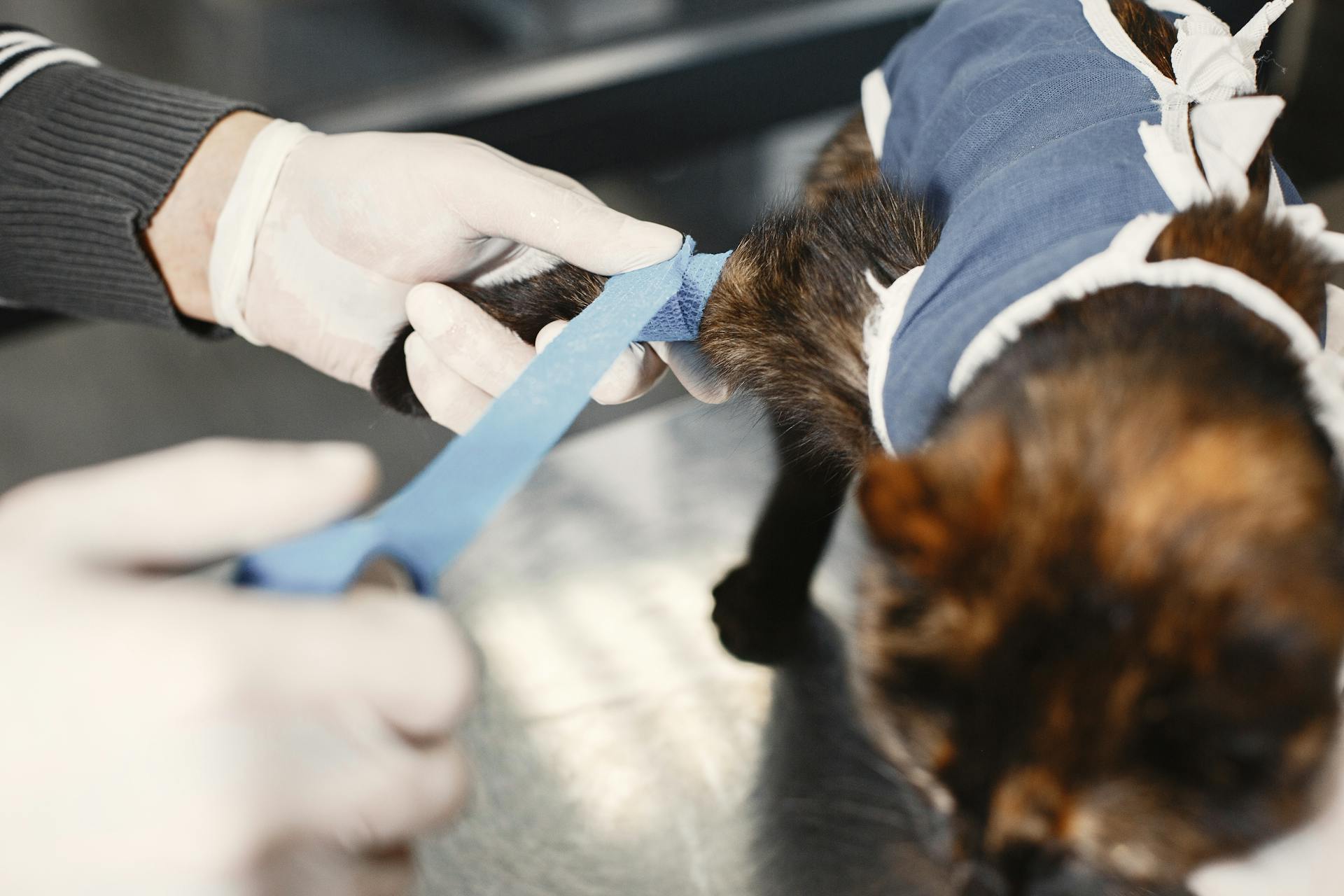
Mammary tumors in rats are a common health issue that can affect any breed or age. They are often benign but can be malignant in some cases.
Rats can develop mammary tumors due to a combination of genetic and environmental factors. Hormonal influences, such as estrogen, play a significant role in the development of mammary tumors.
The likelihood of a rat developing a mammary tumor increases with age, with most cases occurring in rats over the age of 2. Regular veterinary check-ups can help identify tumors early on.
Early detection is key to treating mammary tumors in rats effectively.
Definition and Pathophysiology
Mammary tumors in rats are a complex issue, influenced by multiple factors. Rats with a predisposition to developing mammary tumors include genetic strain, diet, environment, physiologic status, and hormonal status, particularly the presence of prolactin.
Prolactin is secreted during estrus, which occurs every 3 to 5 days in rats, and can stimulate very rapid tumor growth. In addition, the secretion of prolactin increases in both male and female rats as they age.
Rats fed ad libitum have a higher incidence of mammary tumors than those fed restricted amounts of the same diet. This excessive caloric intake can decrease tumor latency and increase tumor growth.
Suggestion: Rat Terrier Growth Chart
Definition
Mammary fibroadenomas are fibrous, benign tumors that arise from glandular tissue.
These tumors are non-cancerous growths that can occur in both female and male rats, although they are more common in females.
Mammary tissue is extensive in rats, covering the ventral, lateral, and dorsal areas of their body, from the shoulder and chin area to the base of the tail.
In male rat fetuses, testosterone is released before mammary tissue development occurs, resulting in rudimentary mammary glands that do not have nipples.
Mammary adenocarcinomas, on the other hand, are malignant growths that arise from glandular organs and are a type of cancer.
Pathophysiology
Rats with a predisposition to developing mammary tumors often have a genetic strain that makes them more susceptible.
Genetic strain is just one factor, diet also plays a significant role in tumor development. Rats fed ad libitum have a higher incidence of mammary tumors compared to those fed restricted amounts of the same diet.
Prolactin levels are also a key factor in mammary tumor growth. Prolactin is secreted during estrus, which occurs every 3 to 5 days in rats, and can stimulate very rapid tumor growth.
As rats age, prolactin secretion increases in both males and females, which can lead to tumor growth. This is why age is a significant factor in the development of mammary tumors in rats.
Rats with pituitary adenomas that secrete prolactin are also at risk of developing mammary tumors. These adenomas can promote chronic stress and obesity, further increasing the risk of tumor development.
Excessive caloric intake can decrease tumor latency and increase tumor growth in rats. This is why feeding rats ad libitum can lead to a higher incidence of tumors, including mammary tumors.
Clinical Presentation and Signs
Mammary tumors in rats can be tricky to spot, but understanding the clinical presentation and signs can help you identify the issue early on.
Rats can develop tumors anywhere from the cervical region to the base of the tail, and these tumors can grow rapidly, becoming very large within weeks.
The tumors may initially appear as a soft, circumscribed growth that can be movable on palpation, but as they grow, they can become more firm and attached.
Rats with mammary tumors may also experience impaired mobility, which can make it difficult for them to move around.
Ulceration and necrosis of tissue are common complications of large mammary tumors, which can lead to blood loss and anemia.
As the tumors grow, they can redirect the rat's dietary intake towards tumor growth, leading to increased appetite with no weight gain.
In later stages, rats with mammary tumors may develop poor appetite, weight loss, and lethargy.
Here are some common clinical signs to look out for:
- Soft, circumscribed (round), or somewhat flat appearing growth that can be movable on palpation
- Impaired mobility
- Ulceration and necrosis of tissue
- Increased appetite with no weight gain
- Poor appetite, weight loss, and lethargy
- Development of one or more growths along mammary chain
Prevention and Etiology
Spaying female rats is a recommended preventative measure against mammary tumors, as it has been documented to prevent or reduce the frequency of tumor development.
Early detection of mammary tumors is crucial, and routine health checks can help identify growths while they are still small. Detection and treatment at this stage can significantly improve outcomes.
Offering a diet that is low in fat, calories, amines, and nitrates can also help prevent mammary tumors in rats.
Prevention
To minimize the risk of mammary tumors in rats, it's essential to take preventative measures. Removing large masses and waiting 3 to 4 weeks before performing an ovariohysterectomy can help minimize surgical time and trauma.
Early ovariohysterectomy or ovariectomy can also lessen the likelihood of mammary tumors by arresting the development of mammary tissue. This is supported by clinical experience, which shows that implementing dietary restrictions, easing environmental stress, and providing exercise and environmental enrichment can help prevent tumor development.
Offering a diet that is low in fat, calories, amines, and nitrates is recommended, as this can help reduce the risk of mammary tumors. Spaying female rats through ovariectomy has also been shown to prevent or reduce the frequency of these tumors.

Routine health checks are crucial in detecting mammary tumors early, allowing for treatment while the growth is still small. This can significantly improve the outcome and reduce operative time and recovery period.
Here are some recommended preventative measures for reducing the risk of mammary tumors in rats:
- Offer a diet low in fat, calories, amines, and nitrates
- Consider spaying female rats through ovariectomy
- Perform routine health checks to detect mammary tumors early
Etiology
Etiology is a crucial aspect of understanding and preventing various conditions.
Genetic predisposition plays a significant role in the development of certain conditions, such as hypertension, which can be inherited from one's parents.
Research suggests that genetics account for approximately 30-50% of the risk of developing hypertension.
Lifestyle factors also contribute to the development of conditions like hypertension, with a diet high in sodium being a significant risk factor.
A diet high in sodium can lead to an increase in blood pressure, which can put a strain on the cardiovascular system.
Environmental factors, such as exposure to air pollution, can also contribute to the development of conditions like asthma.
Air pollution can trigger asthma symptoms, making it essential to take precautions when exposed to polluted air.
Diagnostics and Treatment
Excision and removal of the tumor is the recommended treatment for mammary tumors in rats. This involves surgically removing the tumor, and it's essential to provide the rat with free access to food and water until just before anesthesia.
Pre- or post-op prophylactic broad-spectrum antimicrobials may be indicated in elderly, debilitated, or immunocompromised rats. This is crucial to prevent infection and ensure a smooth recovery.
Rats do experience pain with surgical procedures, so pain medication is necessary. The type of pain medication used post-op should be determined based on the extent of the procedure and the anticipated severity of pain.
For severe pain or the first 24 hours post-op, Buprenex (buprenorphine) or Torbugesic (butorphanol) may be prescribed. For mild to moderate pain, Banamine (flunixin meglumine), Metacam (meloxicam), or carprofen are options.
Diagnostics
Diagnostics can be a crucial step in understanding the health of your rat. Palpation of an ovoid, discoid, or varied shaped mass may be a sign of a tumor.

A large mass may show ulceration or necrosis, which can be a concerning sign.
Rats with tumors may appear cachectic, or thin, as the tumor continues to grow.
Magnetic Resonance Imaging (MRI) can be useful in diagnosing soft tissue tumors, although it may not be available for all small animals.
Histologic examination of tissue is a key diagnostic tool for tumors.
Microscopic examination of an aspirate can also provide valuable information about the tumor.
In some cases, euthanasia may be considered if the tumor significantly affects the rat's quality of life and treatment options have been exhausted.
Treatment
Treatment for mammary tumors in rats involves a combination of surgical excision, pain management, and in some cases, additional medical interventions. Excision and removal of the tumor is the recommended course of action.
Pre-anesthetic fasting is not necessary for rats, as they do not experience vomiting. In fact, it's recommended to provide free access to food and water until just prior to anesthesia.

Pain management is crucial for rats undergoing surgery, and the type of medication used post-op should be determined based on the extent of the procedure and the anticipated severity of pain. Rats do experience pain with surgical procedures.
For severe pain or the first 24 hours post-op, Buprenex (buprenorphine) or Torbugesic (butorphanol) may be prescribed. These medications can provide effective pain relief.
For mild to moderate pain, Banamine (flunixin meglumine), Metacam (meloxicam), or carprofen may be used. However, it's essential to note that these medications should not be used if a corticosteroid has already been prescribed.
If surgery is being performed on a female rat, it's worth discussing with the vet the possibility of also spaying, especially if the rat's general health status is good. Spaying can significantly reduce or remove the hormones that influence mammary tumors.
The table below summarizes some of the pain medication options for rats:
In some cases, additional medical interventions may be necessary, such as using Tamoxifen for malignant estrogen-dependent tumors in elderly rats with high-risk health problems.
Fibroadenomas and Breast Cancer
Fibroadenomas occur more commonly in Sprague-Dawley rats than in other strains, with values as high as 68%. This is significantly higher than the average incidence in other rat strains, which ranges from 20% to 40%.
Fibroadenomas in rats closely resemble their human counterparts, with benign tumors appearing in both species. The cell types present in rat fibroadenomas react with similar immunocytochemical markers as do the human ones.
In rats, fibroadenomas tend to remain confined by the adjacent stroma and show no clear evidence of invasion, making clear distinctions between benign and malignant lesions challenging.
Fibroadenomas in the Breast
Fibroadenomas are a type of non-cancerous breast tumor that can occur in both women and men.
The incidence of fibroadenomas in female rats varies from 20% to 40% in most strains, with some strains like Sprague-Dawley rats having a higher incidence of up to 68%.
In rats, fibroadenomas are more common in certain strains, such as Sprague-Dawley rats, and less common in others, like ACI/N rats.

Fibroadenomas in rats are often found to occur more frequently in females than in males.
The growth of fibroadenomas in rats is influenced by hormones, particularly estrogen, which can stimulate their growth.
Estrogen in combination with gonadotropic hormone can increase the number of successfully transplanted fibroadenomas in rats.
A constant absorption of excessive amounts of estrogen can lead to the formation of fibroadenomas in rats.
Fibroadenomas tend to form in rats during periods of high estrogen levels, such as adolescence, pregnancy, and menopause.
Breast Cancer Molecular Classification
Breast cancer can be classified into several molecular subtypes based on gene expression.
The most well-characterized and widely accepted molecular subtypes are the luminal A, luminal B, HER2/neu, and basal like types.
The basal like subtype commonly displays a triple-negative phenotype.
Luminal A, luminal B, basal-like, and HER2/neu subtypes of ductal carcinoma in situ (DCIS) have also been described.
The basal like subtype is often defined by a lack of estrogen receptor and progesterone receptor protein expression, with an absence of HER2/neu overexpression.
These molecular subtypes are identified through the use of gene expression microarray technology and molecular signatures.
Histopathology and Diagnosis
Rat mammary tumors can be classified into various histological types, including papillomatous, adenomatous, and malignant lesions. These types are similar to those found in humans, but with some specific differences.
The diagnosis of invasive carcinomas in rat mammary glands is based on the presence of unequivocal growth of malignant epithelial cells into the adjacent stroma. This can be challenging due to the diffuse growth of mammary gland ducts into surrounding tissues.
Invasive papillary carcinomas are the most typical and frequent type of tumor in rat mammary glands, often detectable by palpation and characterized by delicate fibrovascular cores and lymphocytic infiltration.
Intriguing read: Types of Dog Tumors with Pictures
Fibroadenomas
Fibroadenomas are a type of benign tumor that can occur in the mammary gland of rats, with a mean incidence ranging from 20% to 40% in most strains.
In some rat strains, like Sprague-Dawley, fibroadenomas occur more commonly, with values as high as 68%. In contrast, ACI/N rats have the lowest incidence, at 4.8%.
The growth of fibroadenomas is influenced by hormones, particularly estrogen. High doses of estrogen can stimulate the growth of pre-existing fibroadenomas, as well as induce hyperplasia in these growths.
Estrogen in combination with gonadotropic hormone can also increase the number of successfully transplanted fibroadenomas in rats. This is consistent with clinical observations that fibroadenomas tend to form during periods of relatively constant estrogenic stimulation, such as adolescence, pregnancy, and menopause.
Histopathology of Gland Carcinoma
The diagnosis of invasive carcinomas in the rat mammary gland is based on the presence of unequivocal growth of malignant epithelial cells into the adjacent stroma.
Invasive carcinomas in the rat mammary gland can be difficult to judge because the mammary gland ducts grow diffusely into the fat pad, adjacent muscle, and subcutaneous tissue of the skin.
The invasive papillary carcinomas are the most typical and frequent type of invasive carcinoma in the rat mammary gland, accounting for 26.05% of all invasive carcinomas.
Papillary carcinomas contain delicate fibrovascular cores, often heavily infiltrated by lymphocytes and mast cells, and can be classified into grade 1 or grade 2 based on the thickness and cytologic characteristics of the epithelium.
For your interest: Perianal Adenoma Female Dog

Papillary carcinomas grade 1 are composed of 1–2 layers of epithelial cells, which emit short epithelial papillae devoid of fibrovascular cores.
Papillary carcinomas grade 2 are formed by papillary projections with sparser fibrovascular cores and solid clusters of epithelial cells, and can occasionally have dilated or cystic luminal spaces.
The earliest change observed in the mammary parenchyma after carcinogen treatment of virgin rats is the dilation of terminal ductal structures, which exhibit thickening of the epithelial lining and can progress to carcinoma in situ.
Experimental Models and Genetic Factors
Experimental models have been used to study mammary tumors in rats, providing valuable insights into the underlying factors that contribute to their development. These models include inducing fibroadenomas using hormones, such as estrogen, which can stimulate the growth of preexisting fibroadenomas.
The growth of fibroadenomas during pregnancy is a notable example of this responsiveness to estrogen. In fact, rats and monkeys can be induced to grow fibroadenomas using high doses of estrogen, and this growth pattern is similar to the rapid mammary growth seen in normal adolescence and early pregnancy.
Genetic factors also play a significant role in the susceptibility of different rat strains to mammary tumorigenesis. For instance, SD and Wistar-Furth rats are the most susceptible to DMBA or NMU mammary tumorigenesis, while Copenhagen rats are essentially completely resistant.
Experimental Induction of Fibroadenomas

Fibroadenomas can be induced in rats using hormones, specifically estrogen. High doses of estrogen have been shown to increase the number of successfully transplanted fibroadenomas in rats.
The rapid growth of preexisting fibroadenomas during pregnancy in humans also suggests a special responsiveness to estrogen. This growth pattern is similar to that of the mammary gland in early pregnancy and adolescence.
Estrogen in combination with gonadotropic hormone has been found to stimulate hyperplasia in fibroadenomas in rats. This combination also increased the number of successfully transplanted fibroadenomas in rats.
Implanting pellets of estrone under the skin of rats has been shown to produce fibroadenomas in the mammary glands. This constant estrogenic stimulation leads to the formation of fibroadenomas, but fluctuations in estrogen levels can induce large cysts instead.
The incidence of fibroadenomas in female rats varies between different strains, with some strains having as high as 68% incidence.
Genetic Factors in Cancer Development
Genetic factors play a significant role in cancer development, particularly in rat mammary tumorigenesis. Research has shown that different rat strains have varying levels of susceptibility to DMBA or NMU-induced mammary tumorigenesis.
The SD and Wistar-Furth rat strains are the most susceptible to DMBA or NMU-induced mammary tumorigenesis, while Copenhagen rats are essentially completely resistant. This suggests that genetic factors can influence an organism's susceptibility to cancer.
In contrast, tumor induction by diethylstilbestrol (DES) is demonstrable in the ACI but not in the SD strain of rats. However, a co-carcinogenic effect of DES with DMBA can be shown in SD rats, indicating that genetic factors can also influence the interaction between different carcinogens.
Transplantation studies have shown that the inherent characteristics of the gland, rather than the host animal, determine the response to DMBA. This suggests that genetic factors within the gland itself play a crucial role in cancer development.
Interestingly, the use of gene expression microarray technology has introduced a new classification of human breast cancer based on molecular signatures. This has allowed for the identification of four distinct molecular subtypes of mammary cells, including the luminal A, luminal B, HER2/neu, and basal-like types.
You might enjoy: Mammary Gland Tumor
Figure 17

The invasive tubular carcinomas in Figure 17 are composed of tubular or alveolar structures. These structures become more irregular in the invasive areas, forming distorted glandular structures.
The areas of invasion are surrounded by a desmoplastic reaction, which is a type of fibrotic response. The epithelial cells in these carcinomas have increased nuclear size.
The nuclei of these epithelial cells contain prominent nucleoli. Stained with H&E, these features can be observed at a magnification of ×40.
Chemical Carcinogens and Biological Importance
Chemical carcinogens like DMBA play a significant role in the development of mammary tumors in rats.
The DMBA rat mammary model has been able to demonstrate that the carcinogen acts on the intermediate cell of the TEB.
The TEB is a crucial structure that evolves to IDP and carcinoma in situ.
A high proliferative activity of the TEB is associated with higher binding of the carcinogen.
The short cell cycle of the TEB makes them less likely to repair the DNA damaged by the carcinogen.
Not all TEBs evolve to carcinomas, and the regulatory mechanism of this process is more complex due to intrinsic properties of the TEB.
IDPs progressing to carcinomas secrete proteoglycans and attract lymphocytes and mast cells.
This highlights the importance of the interaction of the initiated cells with the host as a mechanism in the progression of the disease.
Tables and Figures
In studying mammary tumors in rats, researchers have used various tools to analyze and understand the condition. Table 3 presents a breakdown of the incidence of different types of mammary tumors in rats, with Invasive Papillary Adeno carcinoma Type II being the most common.
The age of the rat is a significant factor in the development of mammary tumors, with the prevalence increasing significantly after 137 weeks of age in female F344 rats. Fibroadenomas, a type of benign tumor, have a peak risk period between 31 and 36 months of age, and their incidence decreases in older rats.
Here are some key structures or lesions that can be confused with mammary tumors, as listed in Table 6:
- Lactating gland: characterized by enlargement of the gland and uniformity affecting all the pairs of glands.
- Salivary gland hyperplasia: located in the submandibular region, with acini containing serous or mucinous type epithelium.
- Clitoridal gland hyperplasia: found in the prepubic location, with uniformly large cells and round, leptochromatic nuclei arranged in acini.
- Lymph nodes: typically normal or reactive, with specific locations and characteristic architecture.
- Abscesses: encapsulated, tense, or fluctuant, with numerous polymorphonuclear leukocytes.
- Skin and adnexal tumors: including basal and squamous cell carcinomas, trichoepithelioma, trichilemmoma, sebaceous adenoma and carcinoma, fibroma, and fibrosarcoma.
- Hibernomas: characterized by typical architecture with numerous fat droplets of varying sizes.
Researchers have also used immunocytochemical evaluation to study mammary tumors in rats, as shown in Table 9, which lists the antibodies used, their providers, and the incubation times.
Table 9
Table 9 is an immunocytochemical evaluation of rat mammary tumors, which is a fancy way of saying it's a table that shows which antibodies were used to test for specific proteins in the tumors.
The table lists three different antibodies: ER-alpha (MC-20), Anti-PR (C-19), and c-erbB-2/Her2/neu Ab-17. These antibodies are used to detect the presence of certain proteins in the tumors.
Here are the details of each antibody:
- ER-alpha (MC-20) is a rabbit polyclonal antibody provided by Santa Cruz, used at a dilution of 1:400.
- Anti-PR (C-19) is a mouse monoclonal antibody provided by Santa Cruz, used at a dilution of 1:800.
- c-erbB-2/Her2/neu Ab-17 is a mouse IgG antibody provided by Thermo Scientific, used at a dilution of 1:1000.
These antibodies are used to test for the presence of specific proteins in the tumors, which can help diagnose the type of tumor and its behavior.
Figure 1
Figure 1 is a crucial element in any table or figure. It's a visual representation of data that helps readers quickly understand complex information.
A well-designed figure can make a significant impact on the reader's understanding of the data. Figures typically include labels, titles, and axes to provide context and clarity.
In Figure 1, the x-axis represents the independent variable, while the y-axis represents the dependent variable. This helps readers see the relationship between the two variables.
The data in Figure 1 is presented in a clear and concise manner, making it easy to read and understand. The use of color and shading adds visual interest and helps to distinguish between different data points.
By including a key or legend in Figure 1, the creator can explain the meaning of the different symbols and colors used in the figure. This helps readers interpret the data correctly.
Figure 19
Figure 19 is a crucial image that helps us understand the characteristics of fibromas and carcinosarcomas. Fibromas are well-circumscribed and non-encapsulated tumors composed of proliferating fibroblasts arranged in interlacing bundles and embedded in variable amounts of collagen fibers.
In some tumors, isolated remnants of glandular epithelium can be found, which suggests that they originate from fibroadenomas. Fibroadenomas are more common in the rat pathology, as we'll discuss in a previous section.
The carcinosarcomas is a rare entity with malignant characteristics in both the epithelium and the stroma. The epithelial component can vary from well-differentiated tubular structures to poorly demarcated and elongated cells.
The specific markers keratin, myoglobin, desmin, and vimentin are useful for separating spindle-shaped epithelial cells from the stromal component. This is a key distinction to make when diagnosing these types of tumors.
Nuclei vary in size and shape, and giant, multinucleated cells may be found in these tumors.
Case Studies and Considerations
The rat mammary tumor model has its limitations, particularly when it comes to studying the metastatic process. Unless the animals are kept for longer periods of time and the methodology for detecting early metastatic events improves, the rat model is not a good model for studying metastasis.
The classification of tumors in the rat model matches well with the criteria used in human pathology, providing an adequate model for understanding the phases of the human disease. The rat model is well suited for studying in situ and invasive lesions.
Immunosurveillance is an area that should be better studied in the rat model, as it could potentially provide new insight into the inflammatory processes involved in the initiation and progression of mammary neoplasia. This could also provide a model for testing vaccines and other tools using cytokine inhibitors.
Case Description
In this case, we have a unique situation where a client's website was hacked, resulting in a loss of sensitive customer data. The hacker demanded a ransom in exchange for the return of the stolen data.
The client's IT department was alerted to the issue and quickly sprang into action to contain the damage. They isolated the affected server to prevent further unauthorized access.
The hacker's demands were met, and the ransom was paid, but not before the hacker had already begun to leak the stolen data online. This led to a significant loss of customer trust and a major reputational hit for the company.
The client's IT department learned a valuable lesson about the importance of regular backups and having a disaster recovery plan in place. They also realized the need for more robust security measures to prevent similar incidents in the future.
Final Considerations
The rat mammary tumor model is well-suited for studying in situ and invasive lesions.
Unless the animals are kept for longer periods of time and the methodology for detecting early metastatic events improves, the rat model is not a good model for studying the metastatic process.
The classification of the tumors matches well with the criteria used in the human pathology, providing an adequate model for understanding these phases of the human disease.
Immunosurveillance is an area that should be better studied, as it could potentially provide new insight into the inflammatory processes involved in the initiation and progression of mammary neoplasia.
The use of immunocytochemical markers allows us to differentiate two cell subtypes, Luminal A and B.
We have not detected any tumors as positive only for HER2/neu or as triple negative.
The presence of histochemical markers does not indicate that the tumors in the rat will behave exactly as they do in the human disease, but will provide an initial point of comparison.
More studies on this subject could be extremely beneficial.
Frequently Asked Questions
How fast do mammary tumors grow in rats?
Mammary tumors in rats can grow rapidly, becoming very large within weeks. Despite their size, rats often appear to tolerate the tumors and continue eating and acting normally.
How long will my rat live with a tumor?
Rats with benign tumors can live a full life after surgery, with some living up to 2 additional years. With proper care and surgery, your rat can enjoy a long and healthy life.
When to euthanize a rat with a tumor?
Euthanasia is considered for rats with tumors that exceed 10% of their body weight, are larger than 1.5 cm, or significantly impact their ability to eat, drink, or move around.
Featured Images: pexels.com