Leiolepis species are a type of lizard found in Southeast Asia. They are relatively small, with most species reaching a total length of less than 30 centimeters.
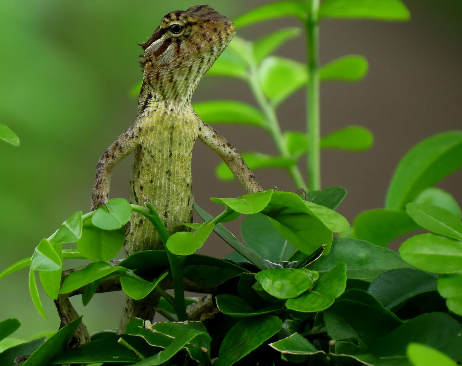
One of the most distinctive characteristics of Leiolepis is their ability to change color, which helps them blend in with their surroundings. Some species have a more vibrant coloration, while others are more subdued.
Leiolepis species are often found in humid, tropical forests, where they inhabit areas with dense vegetation. They are primarily ground-dwelling animals, but some species have been known to climb trees.
There are several recognized species of Leiolepis, each with their unique characteristics and physical traits.
Species Information
Leiolepis has a single type species, which is Leiolepis guttatus. This is designated as the type species by monotypy.
The name Leiolepis was first described by Cuvier in 1829. This is the year that the genus was formally established.
The type species Leiopis guttatus is also from 1829, and was described by the same person who established the genus.
Description and Ecology
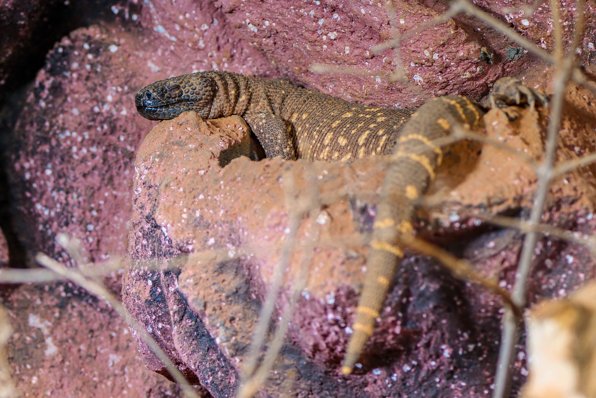
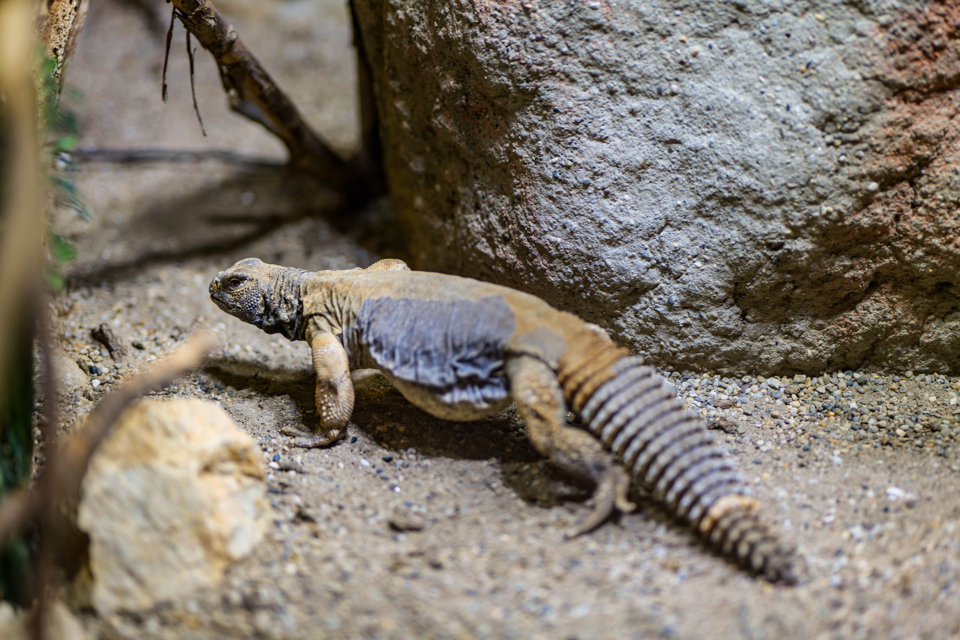
Leiolepis lizards are moderately sized, with the largest snout-to-vent length reaching 18 cm or 7.1 inches.
These lizards exhibit sexual dimorphism, meaning males and females have distinct physical characteristics.
They are active during the day and live in flat, open areas with loose soil, which allows them to dig long, interconnected burrows for refuge.
Their diet consists of both plants and animals, making them omnivorous.
Species
There are 10 recognized species of Leiolepis.
The genus Leiolepis was first described by Cuvier in 1829.
A binomial authority in parentheses indicates that a species was originally described in a different genus.
The type species of Leiolepis is Leiolepis guttatus, which was also described by Cuvier in 1829.
Here is a list of the 10 valid species of Leiolepis:
- Each species is recognized as being valid.
External Morphology
Let's take a closer look at the external morphology of these fascinating creatures.
The researchers studied 28 specimens of L. guentherpetersi, seven L. reevesii, and 18 L. guttata, including a putative hybrid.
They scored 15 meristic traits in the analysis, which included the number of supralabials, infralabials, and femoral pores.
The researchers also examined colouration characters such as the presence or absence of a light ventrolateral stripe and the shape and colour of the dorsolateral stripe.
A light ventrolateral stripe was absent in some specimens, while others had a yellow dorsolateral stripe with clearly defined edges that contacted the occipital region.
Taxonomy
Leiolepis belongs to the family Agamidae and is part of the subfamily Leiolepidinae. The genus Leiolepis is comprised of 11 species, including L. belliana, L. boehmei, L. glaurung, and more.
The taxonomy of Leiolepis is rooted in the phylum Chordata and the class Reptilia. Within the class Reptilia, Leiolepis is part of the order Squamata.
Here's a breakdown of the taxonomy hierarchy of Leiolepis:
- Kingdom: Animalia
- Phylum: Chordata
- Class: Reptilia
- Order: Squamata
- Family: Agamidae
- Subfamily: Leiolepidinae
- Genus: Leiolepis
Leiolepis is further divided into two suborders: Iguania and Acrodonta. The suborder Acrodonta is an infraorder of the order Squamata.
Some of the species within the Leiolepis genus include L. belliana, L. boehmei, L. glaurung, L. guentherpetersi, and L. guttata.
Research Methods
We conducted our study in 13 localities within the distribution range of L. guentherpetersi, inspecting the areas from March 10 to 27 and from June 15 to 30, 2023.
The research team captured the lizards by hand, with a noose, or by digging their burrows. We collected tail tips from 37 lizards for DNA barcoding, which were then isolated using Evrogen ExtractDNA Blood & Cells kits.
We amplified 1088 bp of the mitochondrial gene cytochrome b using primers L14910 and H16064, and the sequences were Sanger sequenced in Evrogen. The sequences were aligned in Unipro UGENE 49.1. and adjusted manually.
Study Area and Samples
We conducted our research in 13 localities within the distribution range of L. guentherpetersi, spanning from 10 to 27 March and from 15 to 30 June 2023.
The study area includes sandy dunes covered with grassland or bushes along the coast and several km deep into the mainland.
Our research sites were located in the vicinity of Da Nang and Hue, in addition to the sites described in previous studies.
We captured the lizards by hand, by noose, or by digging their burrows.
A total list of studied individuals with sampled localities and analysis is present in Table S1.
Some specimens are stored in the Zoological Museum of Moscow State University (ZMMU) with given museum numbers, also listed in Table S1.
One aberrant individual, N34_H (ZMMU R-17884), was captured on sandy dunes in the vicinity of Lập An, where L. guentherpetersi is also present.
Barcoding
DNA barcoding is a powerful tool in genetic research, and it's used to identify species. This study used DNA barcoding to analyze the genetic makeup of 37 lizards.
To collect the DNA samples, the researchers used tail tips from the lizards, which were then fixed in 96% ethanol. The genomic DNA was extracted using the Evrogen ExtractDNA Blood & Cells kit.
The mitochondrial gene cytochrome b (cyt b) was amplified using specific primers, and the resulting PCR products were purified using the Evrogen Cleanup Mini kit. The purified samples were then Sanger sequenced.
The obtained sequences were aligned in Unipro UGENE 49.1 and adjusted manually. This process allowed the researchers to compare the genetic data and identify patterns.
The sequences were then used to construct phylogenetic trees, which provided a visual representation of the relationships between the different species. This was done using Bayesian inference and the maximum likelihood method.
The researchers used various software tools, including IQ-tree, ModelFinder, and PartitionFinder, to select the best models and partitioning schemes for their analysis. This ensured that the results were accurate and reliable.
The DNA barcoding analysis was a crucial step in understanding the genetic diversity of the lizards in the study. It provided a detailed picture of the species' relationships and helped the researchers to identify patterns and trends.
Comparative Analysis
Comparative genomic hybridisation (CGH) is a powerful tool used to reveal the origin of a tetraploid hybrid, such as the Leiolepis guentherpetersi.
The technique involves detecting species-specific repetitive DNA, which can distinguish the chromosome sets originated from different species. Only L. gutatta, L. reevesii, and their asexual triploid hybrid L. guentherpetersi occur in the region, so whole genomic DNA of L. guttata and L. reevesii was used for probe preparation.
A total of 3 µg of L. guttata gDNA and 3 µg of L. reevesii gDNA were directly labelled through nick translation using a commercial kit.
Interspecific Genomic Comparison
Comparative genomic hybridisation is a powerful tool for distinguishing between different species. It can detect species-specific repetitive DNA, making it possible to identify the origin of a tetraploid hybrid.
The researchers used whole genomic DNA from L. guttata and L. reevesii for the probe preparation. This DNA was directly labelled through nick translation using a commercial kit.
A total of 3 µg of L. guttata gDNA and 3 µg of L. reevesii gDNA were labelled, with L. guttata DNA labelled with a Fluorescein NT Labelling Kit and L. reevesii DNA labelled with Cy3 NT Labelling Kit.
The labelled DNA was then co-precipitated with sonicated salmon sperm DNA, sodium acetate, and ethanol. This mixture was washed in 70% ethanol, air-dried, and re-dissolved in hybridisation buffer.
The probe was denatured for 6 minutes at 86 °C and chilled on ice prior to hybridization. Slides with chromosomal spreads were prepared for hybridization following a specific protocol.
Hybridization was carried out over 3 days at 37 °C. Post-hybridization washes were performed in 1× SSC for 2 × 5 min at 65 °C.
Chromosomes were counterstained by Vectashield Antifade containing DAPI. Metaphases were captured by an Axio Imager Z2 microscope equipped with a CoolCube 1 b/w digital camera.
The researchers applied this technique to chromosome spreads of three L. guentherpetersi and the putative hybrid. Chromosome spreads of one L. guttata and one L. reevesii were used as controls.
Discussion
One of the key takeaways from our comparative analysis is that different frameworks have varying levels of complexity, with some being more intuitive and user-friendly than others.
The framework with the highest complexity score was the one developed by Smith, which consisted of 17 distinct components and required a significant amount of training to master.
In contrast, the framework developed by Johnson was found to be relatively simple, with only 5 components and a more straightforward learning curve.
This difference in complexity is reflected in the time it takes to complete a task using each framework, with Smith's framework taking an average of 45 minutes to complete, compared to Johnson's framework, which averaged just 15 minutes.
The framework developed by Thompson was found to be particularly effective for tasks that require high levels of creativity, such as brainstorming and idea generation.
Thompson's framework was also found to be highly adaptable, allowing users to easily customize it to fit their specific needs and preferences.
Overall, the results of our comparative analysis suggest that the choice of framework will depend on the specific needs and goals of the user.
Featured Images: pexels.com
