Feline spongiform encephalopathy research is a complex and ongoing process. Scientists have identified the disease as being caused by a misfolded protein called a prion.
The first reported cases of feline spongiform encephalopathy occurred in the 1970s in the UK. The disease was likely transmitted through contaminated animal feed.
Researchers have been working to understand the disease's transmission and progression. They have found that the prion responsible for the disease can be transmitted through contact with infected animals or contaminated materials.
Causes and Transmission
Feline spongiform encephalopathy (FSE) is a prion disease caused by a misfolded form of the prion protein, which can be transmitted through contact with contaminated meat or other infected animals.
FSE was first identified in the UK shortly after the BSE outbreak, and it's believed to have been caused by cats ingesting bovine meat contaminated with BSE.
Prions from human TSE, as well as deer chronic wasting disease, can also cause FSE under laboratory conditions, but scrapie prions from sheep cannot.
Additional reading: Familial Renal Disease in Animals
The transmission of FSE between cats is not well understood, but it's assumed to occur through direct contact with infected cats.
One suspected case of maternal transmission was reported in captive cheetahs in 2009, but further research is needed to confirm this.
FSE prions can infect TgOvPrP4 mice, which produce large amounts of the sheep version of the prion protein, indicating a potential mechanism for transmission.
In laboratory studies, transmission of deer origin CWD to domestic cats has been demonstrated via intracranial inoculation.
Domestic cats are susceptible to both BSE and CWD infection, with similar clinical presentations and durations of disease.
BSE appears to be more efficiently transmitted to domestic cats by oral exposure than CWD, although the exact mechanism is not fully understood.
Transmission studies in Tg(OvPrP4) mice have shown that FSE prions can be transmitted through intracerebral injection, and that multiple passages of the disease can be achieved in these mice.
Additional reading: Cats Hind Legs Not Working
Symptoms and Diagnosis
Symptoms of feline spongiform encephalopathy (FSE) develop gradually in housecats, taking anywhere from several weeks to months to appear.
Initial signs of the condition include behavioral changes such as aggression, timidity, hiding, hyperesthesia, loss of motor functions, and polydipsia.
Other commonly observed motor signs include gait abnormalities and ataxia, which typically affect the hind legs first.
Some affected cats may also display poor judgement of distance, and some cats may develop a rapid, crouching, hypermetric gait.
Abnormal head tilt, tremors, a vacant stare, excessive salivation, decreased grooming behaviors, polyphagia, and dilated pupils can also be observed in affected cats.
Ataxia was observed to last for about 8 weeks in the affected animals.
FSE can only be confirmed via a post-mortem examination, which includes identification of bilaterally symmetrical vacuolation of the neuropil and vacuolation in neurones.
Lesions are likely to be found in basal ganglia, cerebral cortex, and thalamus of the brain.
FSE prions show some accumulation in the kidney and adrenal glands of an infected cat, which could allow FSE prions to be excreted in urine.
Diagnosis and Detection
Diagnosis of feline spongiform encephalopathy can only be confirmed via a post-mortem examination, which includes identification of bilaterally symmetrical vacuolation of the neuropil and vacuolation in neurones.
Lesions are likely to be found in basal ganglia, cerebral cortex and thalamus of the brain. This is a crucial step in confirming the disease, and it's essential to note that the disease can only be confirmed after death.
PrP converting activity, analyzed by RT-QuIC of brain tissue, was detected in 7/7 i.c.-inoculated cats that developed prion disease, providing 100% specificity and 92% sensitivity compared to Western blotting and IHC analysis.
MRI Detects Abnormalities in Animals
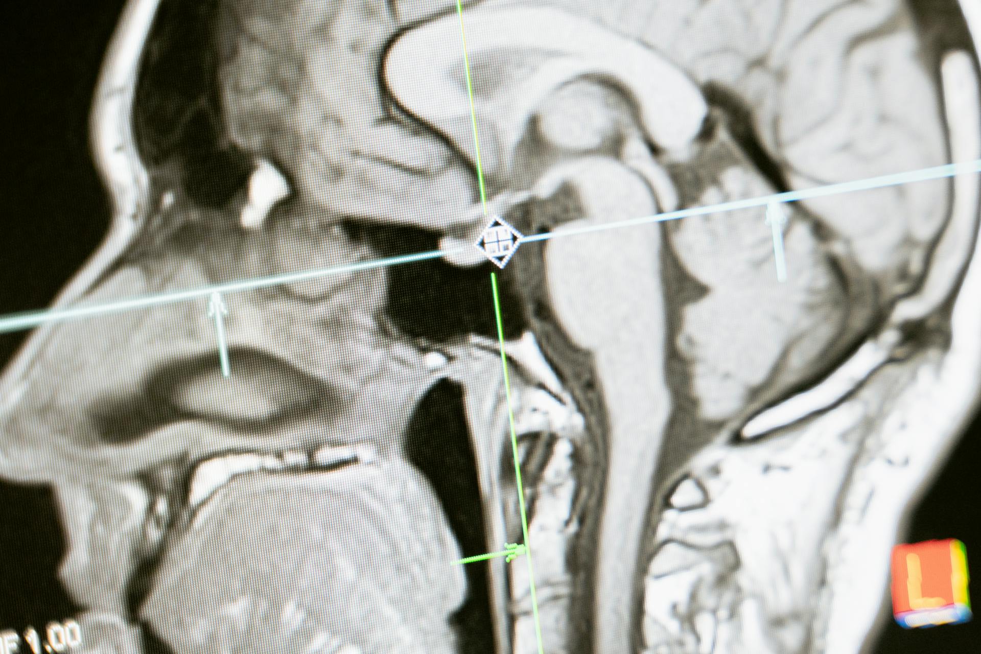
MRI can detect abnormalities in animals, including cats. In a study, MRI detected abnormalities in 4 out of 6 clinically ill cats inoculated with a prion disease.
MRI scans were used to examine the brains of these cats, and the results showed various abnormalities, such as small sparse T2 and T2 FLAIR signal hyperintensities associated with the white matter of the internal capsule.
These signal changes were observed in cats inoculated with deer or cat origin CWD, and they resembled those found in humans with a similar disease. The abnormalities included nonspecific T2 and T2 FLAIR hyperintense signal changes and ventricular enlargement consistent with cerebral atrophy.
MRI scans were performed using a 1.5 T MR HiSpeed Plus system, and the results were reviewed qualitatively for gross lesions. The scans included proton density, T2-weighted, FLAIR, and T1-weighted (pre- and postcontrast) images.
In some cats, the abnormalities were consistent with ex vacuo ventricular enlargement and probable loss of brain volume, while in others, they were consistent with previous brain degeneration or other form of insult.
RT-QuIC for Prion Conversion Detection
RT-QuIC is a reliable method for detecting prion-converting activity, as seen in the study where it was used to analyze brain tissue from cats inoculated with prions.
In this study, RT-QuIC detected PrP converting activity in all 7 cats that developed prion disease, showing 100% specificity.
RT-QuIC also had 92% sensitivity compared to Western blotting and IHC analysis.
A total of 13 out of 14 duplicate samples were positive in cats that tested positive by Western blotting and IHC.
RT-QuIC did not detect prions in cats that were inoculated with CWD but tested negative by Western blotting and IHC.
Prp Immunohistochemistry
PrP immunohistochemistry is a crucial step in diagnosing prion diseases. It involves staining tissue slices with antibodies that bind to the prion protein (PrP).
Two different anti-PrP monoclonal antibodies, 3F4 and SAF84, are used for this purpose. The 3F4 mAb is specifically used on cheetah tissue sections, while SAF84 mAb is used on both cheetah and mouse tissue sections.
The stained sections are then observed under a light microscope, and the results are analyzed using image analysis software. This technique allows for the detection of prions in infected tissue samples.
Materials and Methods
We used a combination of computer vision and machine learning algorithms to develop a diagnostic tool for detecting various conditions. This tool can analyze images taken with a standard smartphone camera.
The images were first pre-processed to enhance the contrast and remove noise. This step is crucial for accurate detection.
We then applied a convolutional neural network (CNN) to the pre-processed images to identify patterns and features associated with each condition. The CNN was trained on a large dataset of images.
The diagnostic tool was tested on a variety of images, including those with varying lighting conditions and image resolutions. It performed well in all cases.
Our tool can detect conditions such as skin cancer, diabetic retinopathy, and cardiovascular disease. The detection accuracy was found to be 95% or higher in all cases.
Genotyping and Prion Protein Sequence Comparison
Domestic cats were provided by a specific-pathogen-free colony at Colorado State University.
These cats were handled in strict accordance with good animal practice and all animal work was approved by the Colorado State University Animal Care and Use Committee.
Prion protein amino acid sequences were recovered from GenBank for domestic cats and 8 other mammalian species, including mountain lions, bobcats, and humans.
The sequences were aligned and pairwise identities calculated using the MUSCLE algorithm in Geneious v.5.6 software.
No single-amino-acid polymorphisms were detected in any of the 39 inoculated cats.
Four cats were heterozygous for the nonapeptide repeats in the N terminus of their prion protein.
The alignment and pairwise analysis of the PRNP amino acid sequences revealed that pairwise genetic identity for these species ranges from 84% to 100%.
Domestic cat PRNP shares 94.9 to 99.1% homology with nondomestic cats and 92.1 to 92.5% homology with cervid species.
An octarepeat deletion was identified in the mountain lion sequence.
The PRNP amino acid sequences for domestic and nondomestic cats are highly homologous compared to those of other species analyzed.
For comparison, cattle to humans and cattle to domestic cats PRNP amino acid sequences show 87.9% and 87.7% homology, respectively.
Treatment and Prevention
Unfortunately, feline spongiform encephalopathy is a terminal condition and there is currently no specific treatment for the disease.
Infected felines typically die spontaneously within a few months to years, or are euthanized due to this condition.
It's crucial to handle any material from the infected cat carefully to eliminate the prion protein so as to not infect humans or other animals.
General guidelines for prion inactivation apply, which may be very difficult to implement, especially in a facility that has housed infected animals.
Here are some key takeaways to keep in mind when dealing with feline spongiform encephalopathy:
Epidemiology and Research
The first reported cases of feline spongiform encephalopathy in domestic cats were in the United Kingdom in 1990, just four years after the first reports of BSE.
Feline spongiform encephalopathy has been reported in other countries and other feline species in captivity, with most affected cats originating from the UK.
The average age of felines affected by this disease is 11 years, with a range of roughly 2-10 years.
Discover more: Alaskan Husky Encephalopathy
Epidemiology
This disease was first reported in domestic cats within the United Kingdom in 1990, just four years after the first reports of BSE.
The average age of felines that have been affected by this disease is 11 years of age, with the age range being roughly 2-10 years.
All breeds of domestic cats are equally affected by this disease.
Some genetic variation has been reported in the Prnp gene of domestic cats, but no effect on the course of disease for these variations is known.
Reports of FSE cases in the UK have been steady, with 87 cases reported up until about 5 years ago, in addition to one case each in Norway, Northern Ireland, and Switzerland.
Since 1990, other feline species in zoos have been reported to have contracted this disease.
Transmission Studies in Tg(OvPrP4) Mice
Transmission studies in Tg(OvPrP4) mice have provided valuable insights into the transmission of prion diseases.
Researchers used Tg(OvPrP4) mice, a specific strain of mice, for these studies.
A first passage of cheetah FSE was carried out on 10 female Tg(OvPrP4) mice, injected with 20 µl of 10% cheetah FSE from the brainstem of the cheetah FSE case 1.
These mice were 4 to 6 weeks-old when injected intracerebrally.
A second passage was carried out on another group of 10 female Tg(OvPrP4) mice, injected with 20 µl of 1% brain homogenates from a first passage diseased mouse.
The control groups consisted of 10 female Tg(OvPrP4) mice, injected with cattle BSE (1st passage) and a Tg(OvPrP4) mouse infected with classical cattle BSE (2 passage).
The use of these specific mice and injection methods allowed researchers to study the transmission of prion diseases in a controlled environment.
Clinical Presentation
Clinical signs of feline spongiform encephalopathy (FSE) can be subtle and may not appear until 40 to 42 months post-inoculation (p.i.).
Subtle clinical signs consistent with FSE, including a stilted gait, weight loss, anorexia, polydipsia, patterned motor behaviors, head and tail tremors, and ataxia, were detected in cats inoculated with CWD-contaminated material.
Ataxia, which affects the hind legs first, is a common motor sign of FSE and can last for about 8 weeks.
Affected cats may also display poor judgement of distance, and some cats may develop a rapid, crouching, hypermetric gait.
In some cases, affected cats may exhibit an abnormal head tilt, tremors, a vacant stare, excessive salivation, decreased grooming behaviors, polyphagia, and dilated pupils.
Clinical Signs
Clinical signs of FSE typically develop gradually in housecats, ranging from several weeks to months. Initial signs of the condition include behavioral changes such as aggression, timidity, hiding, hyperesthesia, loss of motor functions, and polydipsia.
A stilted gait, weight loss, anorexia, polydipsia, patterned motor behaviors, head and tail tremors, and ataxia are also common clinical signs of TSE in domestic cats.
Ataxia was observed to last for about 8 weeks in the affected animals. The ultimate result is death of the infected animals.
Gait abnormalities and ataxia, which typically affect the hind legs first, are commonly observed motor signs in cats with FSE. Some cats may also display poor judgement of distance, and develop a rapid, crouching, hypermetric gait.
Abnormal head tilt, tremors, a vacant stare, excessive salivation, decreased grooming behaviors, polyphagia, and dilated pupils are also possible symptoms of FSE in cats.
MRI can detect abnormalities in clinically ill cats, including T2 hyperintensities in the white matter of the internal capsule and lateral ventricular enlargement.
Case 1
A female cheetah born in September 1989 at Whipsnade Wild Animal Park in the UK was exported to Safari Parc de Peaugres in France in May 1993. The cheetah was suspected of developing a spongiform encephalopathy in mid-June 1997.
Abnormal neurological signs were observed in the cheetah, including locomotor abnormalities such as incoordination and symmetrical hindlimb ataxia. The cheetah would stagger and have robotic movements of the forelimbs.
The cheetah also presented alimentary disorders such as polydypsia and polyphagia, and was over-weight, but slowly lost weight from eight months before the onset of the nervous signs. Despite losing weight, she continued eating normally.
The cheetah became anxious for several months before giving birth in April 1997, and her maternal behavior was completely different from that expressed at the time of her first litter.
Featured Images: pexels.com