A canine gallbladder mucocele is a serious condition that affects dogs, causing a buildup of mucus in the gallbladder, leading to its enlargement.
The condition is often associated with chronic inflammation of the gallbladder, which can be caused by various factors, including biliary tract disease.
Symptoms of a canine gallbladder mucocele can be subtle and may not be immediately apparent, but they can include lethargy, loss of appetite, and vomiting.
In some cases, the mucocele can rupture, leading to severe abdominal pain and potentially life-threatening complications.
Causes and Symptoms
Canine gallbladder mucocele is a serious condition that affects dogs, and understanding its causes and symptoms is crucial for early detection and treatment.
The main cause of a mucocele formation is excessive accumulation of mucus in the gallbladder, and there are several underlying factors that can contribute to this, including gallbladder disease, glucocorticoid therapy, and gallbladder stones.
Some breeds, such as Shetland Sheepdogs and Cocker Spaniels, are more prone to gallbladder mucocele due to their genetic predisposition. Additionally, conditions like endocrine disease, high cholesterol, and pancreatitis can also increase the risk of developing a mucocele.
Dogs with gallbladder mucocele may exhibit a range of symptoms, including lack of appetite, vomiting, abdominal pain, lethargy, and jaundice. If left untreated, the condition can progress to more severe symptoms, such as rapid breathing, rapid heart rate, diarrhea, fever, and excessive thirst and urination.
Here are some common causes of canine gallbladder mucocele:
- Endocrine diseases - hyperadrenocorticism, hypothyroidism, diabetes, etc.
- Hyperlipidemia
- Hypercholesterolemia
- Gallbladder dysmotility
- Mucosal hyperplasia of the gallbladder
Common Causes
Canine gallbladder mucocele, or GBM, is a condition that affects many dogs. The underlying cause of a mucocele formation is unknown, but there are many theories.
Gallbladder disease is one of the possible causes of a mucocele formation. This can lead to a buildup of mucus in the gallbladder, which can cause problems.
Some breeds are more prone to gallbladder mucocele than others. Shetland Sheepdogs and Cocker Spaniels are two breeds that may be more susceptible to this condition.
Dogs with certain medical conditions may be more likely to develop gallbladder mucocele. These conditions include Cushing's disease, diabetes mellitus, or hypothyroidism.
For your interest: Can Allergies Cause Swollen Lymph Nodes in Dogs
A diet high in fat can also contribute to gallbladder mucocele. This is because high fat diets can lead to gallstones or biliary sludge, which can cause problems.
Here are some common causes of canine GBM:
- Endocrine diseases, such as hyperadrenocorticism, hypothyroidism, and diabetes
- Hyperlipidemia
- Hypercholesterolemia
- Gallbladder dysmotility
- Mucosal hyperplasia of the gallbladder
These conditions can all contribute to the development of gallbladder mucocele in dogs.
Who Gets a Condition?
Dogs of any sex can develop gallbladder mucoceles, but geriatric patients are commonly affected.
The median age for dogs with this condition is 10 years, with a range of 3.5-15.0 years.
Dogs weighing less than 20 kilograms (44 pounds) are over-represented, making up more than 70% of reported patients.
Certain breeds are more likely to develop gallbladder mucoceles. Here are a few examples:
- Shetland Sheepdogs
- Miniature Schnauzers
- Cocker Spaniels
Diagnosis and Treatment
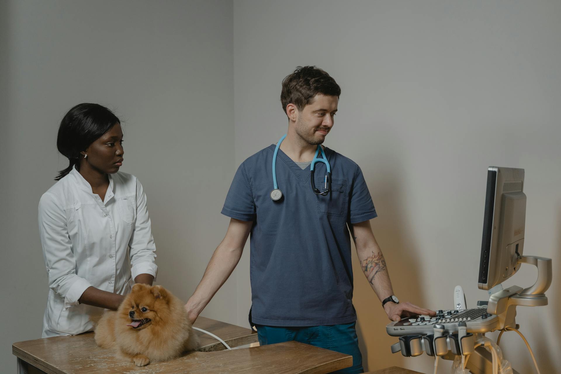
Diagnosis of canine gallbladder mucocele typically involves a physical exam, blood work, and abdominal ultrasound. An ultrasound can detect a mucocele and help differentiate it from biliary sludge.
Your veterinarian may perform a cholecystocentesis, where they use ultrasound-guided laparoscopy to collect gallbladder bile for testing. This can help diagnose bacterial infections and other complications.
For another approach, see: Skin Relief for Dogs with Allergies
Blood tests, including a CBC, blood chemistry test, and blood gas test, can evaluate your pet's overall physical condition and check for signs of inflammation, liver abnormalities, and hypercholesterolemia.
Abdominal radiographs, or X-rays, can provide a general perspective of the organs in the abdominal cavity and help diagnose a ruptured gallbladder.
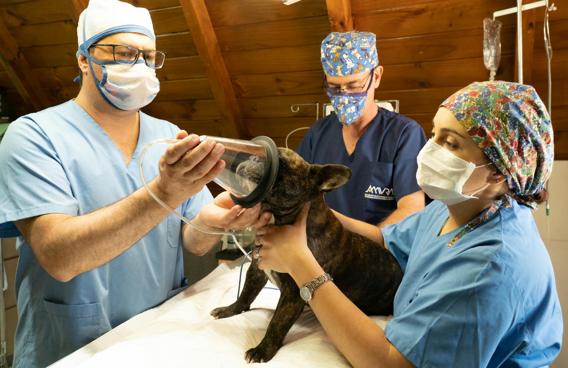
If your dog is diagnosed with a gallbladder mucocele, treatment may involve medical management or surgery. Medical management may include antibiotics, choleretics, and hepatoprotectants to stimulate bile excretion and protect the liver.
Here are some common treatment options for canine gallbladder mucocele:
Surgery is usually recommended for dogs with a ruptured gallbladder, biliary duct obstruction, or severe symptoms. The surgery involves removing the gallbladder and flushing the abdominal cavity clean.
After surgery, your dog may require antibiotics, antiemetics, and pain medications to manage pain and prevent complications. It's essential to follow your veterinarian's instructions and monitor your dog's condition closely during the recovery period.
Here's an interesting read: English Bulldog Cherry Eye Surgery Cost
Anatomy and Pathophysiology
The gallbladder is an excretory organ found between the quadrate and right medial liver lobes. It's pear-shaped and composed of a fundus, body, and neck.
The gallbladder wall has five layers: epithelium, mucosa, tunica muscularis externa, tunica serosa, and tunica adventitia. The epithelium is simple columnar and highly absorptive, playing an important role in gallbladder function by secreting mucin, immunoglobulins, and acid.
The tunica serosa is a membranous layer surrounding the gallbladder that faces away from the liver, while the tunica adventitia is the outermost gallbladder layer and faces the liver.
Pathophysiology
Before we dive into the pathophysiology of a gallbladder mucocele, it's essential to understand what a mucocele is. A gallbladder mucocele is an abnormally distended gallbladder containing a buildup of luminal mucus.
The gallbladder appears enlarged and contains gelatinous material, which can be a mucus cast or inspissated bile. In fact, before 2000, mucoceles were considered rare and were often noted as incidental necropsy findings.
See what others are reading: Kennel Cough Mucus
Predisposition to mucoceles may be associated with dyslipidemias in particular breeds such as Shetland sheepdogs. This is a breed-specific condition that requires attention.
Mucocele formation is not directly associated with extrahepatic biliary duct obstruction in dogs, but rather the opposite: obstruction is secondary to mucocele formation. This is an important distinction for veterinarians and pet owners to understand.
The most widely supported theory of canine mucocele formation implicates mucus-secreting cell proliferation and dysfunction. In this condition, cystic mucinous hyperplasia of the gallbladder epithelium occurs, and the gallbladder epithelial cells secrete excessive mucus into the gallbladder lumen.
Biliary sludge, a mixture of precipitated cholesterol crystals, bile pigments, bile salts, and mucin, may be associated with disease in dogs but is also often seen in clinically normal geriatric dogs. The significance of biliary sludge in dogs is currently unknown.
Anatomy
The gallbladder is a pear-shaped organ located between the quadrate and right medial liver lobes. It's a pretty interesting structure, and understanding its anatomy is key to grasping its function.
The gallbladder is made up of a fundus, body, and neck, which all work together to store and release bile into the digestive system.
The hepatic ducts join the cystic duct to form the common bile duct, which leads to the duodenum, a crucial part of the digestive process.
The gallbladder wall has five distinct layers: epithelium, mucosa, tunica muscularis externa, tunica serosa, and tunica adventitia. This complex structure allows the gallbladder to perform its vital functions.
The epithelium, which is the innermost layer, is simple columnar and highly absorptive, playing a key role in gallbladder function by secreting mucin, immunoglobulins, and acid.
The tunica serosa, a membranous layer, surrounds the gallbladder and faces away from the liver, providing a protective barrier.
The tunica adventitia, the outermost layer, faces the liver and completes the gallbladder's anatomy.
Clinical Presentation
Clinical illness with canine gallbladder mucocele averages ~5 days, although some dogs have vague episodic clinical signs for months. Dogs with gallbladder mucocele may not show any clinical signs at all.
Intriguing read: Signs of Allergies in Dogs
Vomiting is the most common clinical sign, occurring in 77% of affected dogs. Lethargy, loss of appetite, and abdominal discomfort are also common symptoms.
Jaundice or yellowing of the skin is a more serious symptom, occurring in 47% of cases. Abdominal distention, diarrhea, fever, and rapid breathing are also possible symptoms.
The initial symptoms are often vague and intermittent, such as loss of appetite and abdominal discomfort. As the disease progresses, more noticeable symptoms like jaundice, vomiting, and diarrhea may occur.
Common clinical signs include anorexia, abdominal discomfort, vomiting, lethargy, icterus or yellowing of the skin, diarrhea, fever, and abdominal distention.
Here are the top clinical signs of canine gallbladder mucocele:
- Vomiting (77%)
- Lethargy (73%)
- Loss of appetite (71%)
- Icterus or yellowing of the skin (47%)
- Abdominal discomfort (44%)
- Diarrhea (29%)
- Fever (20%)
Veterinary Care
Regular veterinary check-ups are crucial in detecting canine gallbladder mucocele early on.
A gallbladder mucocele can be difficult to diagnose, but your veterinarian may suspect it based on your dog's symptoms and medical history.
Symptoms of a gallbladder mucocele can include vomiting, diarrhea, and abdominal pain.
Dogs with a gallbladder mucocele may also appear lethargic and lose their appetite.
In some cases, a gallbladder mucocele can rupture, leading to severe abdominal pain and potentially life-threatening complications.
Your veterinarian may recommend surgery to remove the gallbladder and alleviate symptoms.
In some cases, surgery may not be possible, and your veterinarian may recommend a more conservative approach.
The prognosis for dogs with a gallbladder mucocele depends on the severity of the condition and the overall health of the dog.
With proper veterinary care, some dogs with a gallbladder mucocele can recover fully.
However, in severe cases, the condition can be fatal.
Regular veterinary check-ups can help prevent gallbladder mucocele by detecting underlying conditions that may contribute to its development.
Your veterinarian can provide guidance on how to care for your dog and prevent complications.
Intriguing read: How to Prevent Canine Parvovirus
Frequently Asked Questions
Can a dog survive gallbladder mucocele?
Survival rates for dogs with gallbladder mucocele vary, but early surgical intervention can significantly improve chances of survival. Mortality rates are reported to be between 20-39%
Can a gallbladder mucocele go away without surgery?
In some mild cases, a gallbladder mucocele may resolve with medical management, but surgery is often necessary to prevent complications. Medical treatment with ursodiol may help improve bile flow, but its effectiveness varies.
Sources
- https://www.merckvetmanual.com/digestive-system/hepatic-diseases-of-small-animals/canine-gallbladder-mucocele
- https://wagwalking.com/condition/canine-gallbladder-mucocele
- https://www.dvm360.com/view/update-gallbladder-mucoceles-dogs
- https://criticalcaredvm.com/gallbladder-mucoceles-dogs/
- https://www.buddydoc.io/blog/gallbladder-mucocele-in-dogs-causes-and-symptoms-of-canine-gbm
Featured Images: pexels.com